What Are Venous Compression Syndromes and Why Should You Care?
- surgeonslab1
- May 16, 2025
- 4 min read
Venous compression syndromes represent a complex category of vascular conditions where external anatomical structures impinge on veins, compromising blood flow and leading to venous hypertension. While these conditions may initially present subtly, their progressive nature can significantly impact patients' quality of life when left unaddressed.
This article explores the pathophysiology, clinical presentations, diagnostic approaches, and contemporary treatment strategies for the most common venous compression syndromes.
The Pathophysiological Basis
At their core, venous compression syndromes develop when rigid anatomical structures, typically arteries, bones, or ligaments, apply external pressure on adjacent veins. This mechanical compression creates a bottleneck effect, increasing upstream venous pressure and impeding normal blood return.
Over time, chronic compression induces secondary changes, including intimal hypertrophy, fibrous spurs, and, in severe cases, thrombosis. The resulting venous hypertension manifests as pain, swelling, and varicosities and potentially progresses to venous ulceration or organ dysfunction if intervention is delayed.
May-Thurner Syndrome: The Classic Iliac Compression
May-Thurner syndrome (MTS), the most widely recognized venous compression disorder, involves the left common iliac vein being compressed by the overlying right common iliac artery against the fifth lumbar vertebra. This anatomical configuration predominantly affects women between 20-50 years of age, manifesting as left-sided lower extremity edema and heaviness that worsens with prolonged standing or activity. More concerning presentations include recurrent deep vein thrombosis (DVT) despite adequate anticoagulation therapy.
The chronic compression and flow turbulence within the iliac vein can lead to endothelial injury, development of venous spurs, and progressive stenosis. As the condition advances, patients may develop extensive pelvic collateral vessels that attempt to bypass the obstruction, a compensatory mechanism that often proves insufficient as symptoms progress.
Nutcracker Syndrome: Renal Vein Entrapment
When the left renal vein becomes compressed between the superior mesenteric artery and the abdominal aorta, Nutcracker syndrome emerges. This condition presents with a distinctive constellation of symptoms, including microscopic or gross hematuria, orthostatic proteinuria (especially in younger patients), and flank pain that may radiate to the inner thigh.
Female patients often experience additional symptoms of pelvic congestion, chronic pelvic pain, dyspareunia, and vulvar or perianal varicosities due to reflux into ovarian veins when renal vein hypertension develops. Left untreated, this condition can lead to renal venous hypertension, potentially compromising kidney function over time.
Thoracic Outlet Syndrome and Paget-Schroetter Syndrome
At the thoracic outlet, venous compression presents as Paget-Schroetter syndrome, where the subclavian vein becomes compressed between the first rib and clavicle.
Unlike other compression syndromes, this condition is often precipitated by repetitive arm movements, particularly in young athletes engaged in overhead activities such as swimming, baseball, or weightlifting. The acute presentation typically includes sudden-onset arm swelling, cyanosis, and visible collateral veins traversing the shoulder and chest wall.
Modern Diagnostic Approaches
The diagnosis of venous compression syndromes has evolved significantly with advances in imaging technology. While Doppler ultrasound serves as a valuable first-line screening tool to assess flow dynamics and visualize collateral formations, cross-sectional imaging provides more definitive anatomical information.
CT venography or MR venography offers a high-resolution visualization of vessel anatomy and the precise point of compression while also revealing the extent of collateral development. For definitive assessment, catheter venography combined with pressure measurements remains the gold standard, confirming significant pressure gradients across the lesion (typically >2 mm Hg).
Intravascular ultrasound (IVUS) has emerged as an invaluable adjunct, providing direct luminal imaging to quantify stenosis severity and guide intervention planning.
Contemporary Treatment Strategies
Management approaches to venous compression syndromes have shifted dramatically toward minimally invasive endovascular techniques. While conservative measures such as compression stockings and leg elevation may alleviate symptoms in mild cases, persistent or severe presentations typically warrant intervention.
Endovascular stenting has become the first-line therapy for symptomatic May-Thurner and Nutcracker syndromes, with dedicated venous stents demonstrating primary patency rates exceeding 90% at one year. The procedure involves balloon angioplasty to pre-dilate the narrowed segment, followed by the deployment of a self-expanding stent designed to withstand external compression forces.
For patients with anatomical configurations unsuitable for stenting or following endovascular failures, surgical options remain viable. These include transposition of the crossing artery or vein, creation of venous bypass grafts, or, in cases of Nutcracker syndrome, nephropexy to relieve the angular compression of the renal vein.
Long-term Management and Outcomes
Post-intervention surveillance is critical to ensuring sustained treatment success. Contemporary protocols recommend duplex ultrasound evaluations at 3, 6, and 12 months following intervention, with annual assessments thereafter. When properly diagnosed and treated, most patients experience significant symptom relief. Studies report resolution of hematuria in approximately 80% of Nutcracker cases and substantial improvement in leg symptoms in May-Thurner patients.
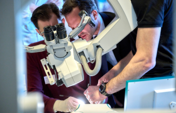
Enhancing Procedural Competence Through Simulation
The technical complexity of endovascular interventions for venous compression syndromes necessitates specialized training. Simulation platforms like SurgeonsLab's Venous Model offer clinicians risk-free environments to master catheter navigation, stenting techniques, and management of potential complications before performing procedures on patients. These high-fidelity training modalities represent an important advancement in procedural education, potentially reducing complications and improving patient outcomes.
With increased awareness among clinicians, advanced diagnostic capabilities, and evolving treatment options, patients with venous compression syndromes can now access effective therapies that restore venous outflow and prevent the long-term sequelae of chronic venous hypertension.