Innovative Training Solutions for MMA Embolization Procedures
- surgeonslab1
- Feb 6, 2025
- 4 min read
For chronic subdural hematomas (cSDH), middle meningeal artery (MMA) embolization has become a novel, less invasive therapy that presents an alluring substitute for conventional surgical techniques. MMA ablation helps prevent repeated bleeds by blocking the blood flow to weak blood vessels around the hematoma, which can have high rates of problems with open surgery.
But what is MMA, and in light of embolization, how does it work? Furthermore, how can creative training tools such as advanced simulation models provide surgeons with what's needed to carry out these difficult operations safely and effectively? This piece addresses important issues by delving deeply into MMA embolization and its training.
What Is MMA and Why Is It Important?
"MMA" stands for the middle meningeal artery, which is a blood vessel that takes blood to the dura mater (the outer meningeal layer) and some parts of the skull. Beginning in the infratemporal fossa as a branch of the maxillary artery, the MMA separates into anterior and posterior branches after passing via the foramen spinosum into the skull. Its anatomical path renders it more prone to damage; for example, the usual cause of epidural hematoma is a rupture of the anterior branch under the pterion.
Beyond catastrophic injuries, the development of cSDH is much aided by the MMA. From delicate, neo-vascularized membranes forming on the outside of the hematoma, a cycle of inflammation and microbleeds results in chronic subdural hematoma. The MMA becomes the ideal target for embolization by supplying the blood to these diseased membranes. Reducing future bleeding, helping hematoma clearance, and finally lowering recurrence rates may all be achieved by closing this artery.
MMA Embolization: Procedure and Technical Aspects
MMA embolization is done under moderate sedation or general anesthesia. It involves several key steps:
Vascular Access and Catheterization
To access the MMA, a catheter is passed under real-time imaging direction from the femoral or radial artery. Given the complex vasography and the tiny size of the target arteries, this stage calls for exact catheter manipulation.
Selective Angiography
Selective angiography is done after the microcatheter is in situ to map the MMA's branches and find the arteries supplying the outer membrane of the hematoma. Planning the embolic agent injection depends on this imaging stage.
Injection of Embolic Material
A liquid embolic agent, like n-butyl cyanoacrylate (n-BCA) or Onyx®or micro-embolization coils like SwiftPAC or OPTIMA, are introduced. These agents guarantee that they reach the distal neo capillary network and enable exact imaging of their passage by being radio-opaque. Known as the "MMA Embolic" treatment, the operation treats the fundamental cause of hematoma growth by blocking these pathways.
Procedure Completion and Recovery
The catcher is blocked after embolization, closing the artery puncture hole. Most recently, MMA embolization results in a quicker recovery period and fewer difficulties than open surgical evacuation.
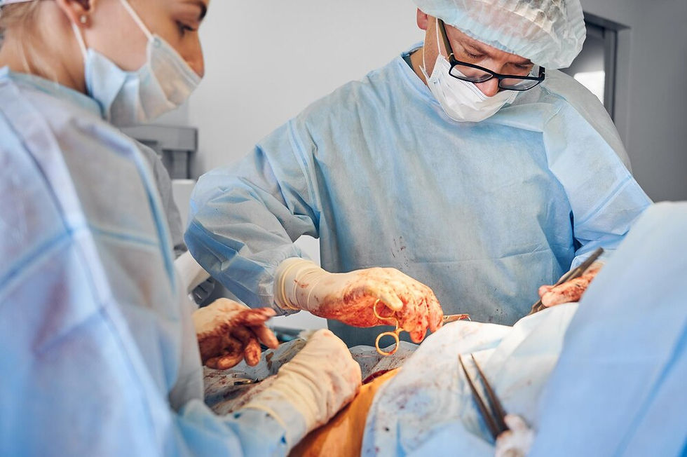
The Need for Advanced Training in MMA Embolization
Performing MMA emoblization needs very skilled techniques. Navigating microcatheters through a complicated vascular network and managing the injection of liquid embolic agents requires great accuracy; even little mistakes may cause major problems, including inadvertent embolization of collateral arteries serving important tissues.
Particularly for unusual or complicated situations, training in neurointerventions has depended on apprenticeship models that may not provide enough chances for the experience. By providing a secure, regulated environment free of patient risk for skill development and refinement, innovative simulation-based training closes this gap.
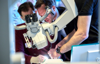
Innovative Simulation-Based Training Solutions
Physical and 3D-Printed Models
Using realistic models created by 3D printing is one of the most exciting developments in education. For instance, SurgeonsLab offers a sophisticated MMA model that faithfully reproduces the extracranial middle meningeal artery in both 2D and 3D versions. These models have:
Customizable Anatomy: The models integrate realistic microvasculature (down to sizes as tiny as 0.2 mm) and extended arterial branches, therefore allowing trainees to gain a thorough awareness of vascular paths and microcatheter navigation.
Perfusion Closed-Loop System: These simulations let practitioners see the consequences of embolic agent injection in a dynamic, life-like environment by emulating genuine blood flow dynamics.
Angiographic and Optical Navigation Compatibility: These models, which are designed to fit imaging modalities well, offer a training experience that mirrors real-world clinical operations.
The Role of SurgeonsLab in Advancing MMA Embolization Training
SurgeonsLab is one of the leading providers of innovative neurovascular training solutions; it has developed an advanced Middle Meningeal Artery embolization model for better procedural accuracy and skill development. Interventional neuro radiologists can practice embolization techniques using accurate anatomy models. This helps them gain skills and confidence before performing these procedures on patients.
To Conclude
MMA embolization is changing how we treat chronic subdural hematomas. It is a better, less invasive option than standard surgery. To learn this method, you need clear instructions and practical experience. Medical education is being transformed by innovative thoughts such as 3D-printed anatomical models, VR-based simulations, and high-fidelity training tools developed via SurgeonsLab, which guarantees that surgeons are confident to execute these challenging operations.
As technology improves, practice training will become more important for better embolization methods, lowering problems, and improving patient care. Using these innovative technologies will provide the next generation of neuro-interventionalists with the knowledge required to maximize results in MMA embolization operations.